Foundation and multimodal models
Vision-language models that read an image and a report together, because clinicians never look at either in isolation.
I work at the intersection of AI and generative AI for radiology, medical imaging, developing research systems that connect images, language, and clinical data for applications in oncology 🫁 🧠 and cardiology 🫀.
Working on medical imaging, multimodal AI, or clinically grounded generative models? Let's grab a coffee ☕ together.
My work sits where engineering, radiology, and clinical research overlap. Cardiac MRI, cancer CT and MR imaging, generative models, and the careful translation from an experiment into something a clinician would trust.
Vision-language models that read an image and a report together, because clinicians never look at either in isolation.
Latent diffusion for medical images, and the harder question of when synthetic data actually helps.
Diagnosis, risk stratification, and treatment response, evaluated the way a clinical question would be.
2026
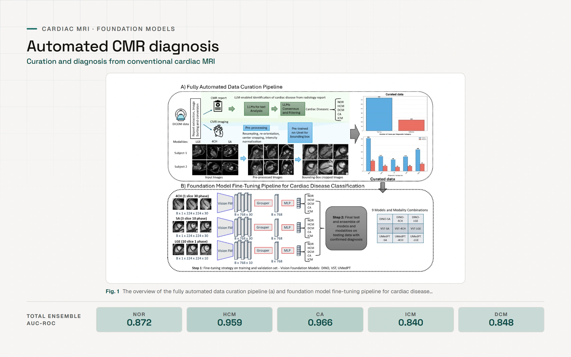
Cardiovascular magnetic resonance (CMR) imaging enables non-invasive assessment of myocardial structure, function, and pathology, but requires substantial experience in interpretation of CMR images that could be supported by artificial intelligence (AI)-based models. We developed an automated data curation pipeline using locally-run large language models to extract diagnostic labels from CMR reports, then fine-tuned three vision foundation models (DINO, VST, UMedPT) on multimodal imaging data across 988 curated cases. The ensemble approach achieved diagnostic performance with AUC-ROC values up to 0.966 for cardiac amyloidosis and 0.959 for hypertrophic cardiomyopathy. Training code and model weights are publicly available.
2025
We investigated the utilization of privacy-preserving, locally-deployed, open-source large language models to extract diagnostic information from free-text cardiovascular magnetic resonance (CMR) reports. Nine open-source LLMs were evaluated on their ability to identify diagnoses and classify patients into cardiac diagnostic categories based on descriptive findings in 109 clinical CMR reports, scored with accuracy, precision, recall, and F1, with confusion matrices used to examine misclassification patterns. Most models performed exceptionally well: Google's Gemma2 achieved the highest average F1 score of 0.98, followed by Qwen2.5:32B and DeepseekR1-32B at 0.96 and 0.95. All other evaluated models scored above 0.93, apart from Mistral and DeepseekR1-7B. The top four LLMs outperformed a board-certified cardiologist (F1 score of 0.94) across every evaluation metric. These findings demonstrate the feasibility of using open-source, privacy-preserving LLMs in clinical settings for automated, accurate, fast, and resource-efficient diagnostic categorization of imaging reports.
2025
Radiomics could play a relevant role in precision oncology by offering precise quantitative support for clinical decision-making. The Radiomics Quality Score (RQS), developed to assess the rigour of radiomics studies, has been widely adopted by researchers since its 1.0 benchmark, but an updated framework is needed to account for evolving knowledge around fairness, explainability, quality control, and harmonization. This review introduces RQS 2.0, which keeps the scientific rigour of its predecessor while addressing these contemporary needs to potentially accelerate clinical translation. It also introduces radiomics readiness levels, inspired by the technology readiness level framework and integrated into RQS 2.0, reflecting nine distinct levels of incremental improvement toward clinical implementation, alongside a strategic vision for precision oncology's future direction.
2025
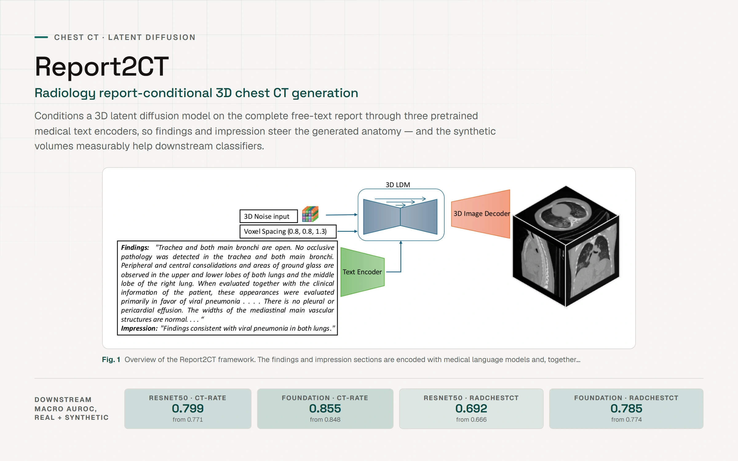
Text-to-image latent diffusion models have advanced medical image synthesis, but 3D CT generation remains limited, and existing approaches rely on simplified prompts that neglect the rich semantic detail in full radiology reports. Report2CT is a radiology report-conditional latent diffusion framework that synthesizes 3D chest CT volumes directly from free-text reports, integrating three pretrained medical text encoders (BiomedVLP CXR BERT, MedEmbed, and ClinicalBERT) to capture nuanced clinical context. The model was trained on 20,000 CT volumes from the CT-RATE dataset, conditioned on complete reports and voxel spacing. Evaluated with Frechet Inception Distance and CLIP-based semantic alignment against the GenerateCT baseline, Report2CT generated anatomically consistent volumes with strong text-image alignment; multi-encoder conditioning improved CLIP scores, and classifier-free guidance further enhanced alignment with only a minor FID trade-off. Report2CT ranked first in the VLM3D Challenge at MICCAI 2025 on Text-Conditional CT Generation, achieving state-of-the-art performance across all evaluation metrics.